<img height="1" width="1" style="display:none;" alt="" src="https://dc.ads.linkedin.com/collect/?pid=54935&amp;fmt=gif">


10th Annual
Practical Radiology – Core Topics in Emergency Radiology

Radiology CME Conference 


February 29 - March 2, 2024 | ARIA Resort & Casino, Las Vegas

17.00 AMA PRA Category 1 Credit(s)™

Open to all radiologists!


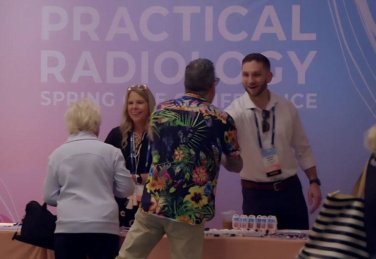
Thank you to those who attended our 2024 CME conference! 

"This was the best radiology conference

I’ve ever attended in my career. Great content, great people."

- 2023 Conference Attendee


2024 Conference Speakers


Benjamin W. Strong, MD
Chief Medical Officer, vRad
Dr. Strong joined vRad as a teleradiologist in July 2004, and served as a medical director within vRad’s practice from October 2005 to October 2012. Dr. Strong is board certified in both radiology and internal medicine. Prior to joining vRad, Dr. Strong worked as an emergency room physician for the IHS, as a private practice radiologist for Inland Imaging in Spokane, WA, and as a Clinical Assistant Professor of Radiology for the University of Arizona Health Sciences Center.
Fabio Paes, MD
Associate Professor of Diagnostic Radiology, University of Miami - Miller School of Medicine
Dr. Paes completed his internship, Diagnostic Radiology and Nuclear Medicine residency at University of Miami/Jackson Memorial Hospital, becoming board certified in Diagnostic Radiology in 2013, and obtained his MBA degree in Healthcare Administration at Florida International University in 2016. Dr. Paes is a full-time faculty in the ER/Trauma Imaging Division, having an active role in the education and training of residents and fellows. 
David Naeger, MD, FAUR
Director of Radiology, Denver Health; Professor and Vice Chair of Radiology, University of Colorado School of Medicine
Dr. Naeger completed his residency at University of California-San Francisco (UCSF) in Diagnostic Radiology, during which he served as a chief resident and was awarded the prestigious Elmer Ng Award. At UCSF, he also completed a research fellowship and a clinical fellowship in Thoracic Imaging and Nuclear Medicine. After his training, he was appointed to the faculty at UCSF, ultimately serving as the Department of Radiology's Associate Chair for Education. In 2019, Dr. Naeger joined Denver Health where he has oversight of Denver Health’s 160-person team, including radiologists, administrators, technologists, nurses, transporters, clerks, and medical physics professionals.
Kathryn Fowler, MD
Diagnostic Radiologist/ Chief, Body Imaging Section/ Professor of Radiology, UC San Diego Health
Kathryn Fowler, MD, is a board-certified radiologist who specializes in body and oncological imaging. As a professor in the Department of Radiology, Dr. Fowler is involved in the teaching of fellows, residents and medical students at the UC San Diego School of Medicine, where she also serves as director of abdominal and pelvic magnetic resonance imaging (MRI) and director of the Liver Imaging Group (LIG). She is also a T32 research training grant director. She has lectured nationally and internationally about liver cancer, transplantation, surgery, imaging and MRI, and written several book chapters and over 150 peer-reviewed publications.
Unni Udayasankar, MD
Professor/ Vice Chair of Quality and Safety Program/ Director of Neuroradiology Fellowship Program, University of Arizona College of Medicine Tucson

Dr. Udayasankar received his radiology training from Emory University School of Medicine including Pediatric Radiology and Neuroradiology fellowships. After his training he worked as a pediatric neuroradiologist at Cleveland Clinic Hospital from 2010-2015. Currently he also serves as the program director for Neuroradiology fellowship at the University of Arizona. He has an extensive academic credentials including research, education and clinical expertise in the area of pediatric neuroradiology and pediatric radiology. One of his key research interests is utilization of faster imaging techniques in the area of emergency neural matching. 

Brian Baker
Senior Director of Software Engineering/Machine Learning and DevOps, vRad
Mr. Baker is committed to enhancing patient care through process improvement and technological innovation. He leads the vRad team responsible for the industry’s most advanced radiological workflow system, including the design, development and integration of leading-edge AI applications. He promotes a collaborative culture as the foundation of these systems that help radiologists provide diagnostic insights more efficiently and effectively every day.
David L. Smith, MD
Professor of Clinical Radiology/ Co-Director of Musculoskeletal Research, LSU Health
Dr. Smith graduated from Wake Forest University's Radiology residency program (2009) and from the MRI fellowship at the Ochsner Clinic Foundation in New Orleans (2010). He practices, teaches, and lectures in Cardiothoracic, Abdominopelvic, and Musculoskeletal subspecialties. He is a member of several professional and academic societies and committees and has lectured nationally and internationally on a range of topics in Radiology, including his area of special interest, Interstitial Lung Disease. Dr. Smith is the Director of Medical Student Education in Radiology at LSU Health New Orleans and teaches medical students, Radiology residents and fellows, and residents and fellows from a number of other specialties daily. 
Bharti Khurana, MD, MBA, FACR, FASER
Founder and Director, Trauma Imaging Research and Innovation Center/ Associate Professor, Harvard Medical School
Dr. Khurana is an associate professor of radiology at Harvard Medical School, an emergency radiologist at the Brigham and Women’s Hospital, and the founding director of the Trauma Imaging Research and Innovation Center (TIRIC). Her clinical and research efforts involve accurate and meaningful imaging interpretation with rapid application of clinical knowledge for patients in the emergency department. She is the director of the Intensive Review of Emergency Radiology CME course at Harvard Medical School and a research faculty in the Department of Medicine at Brigham and Women’s Hospital. She is the author of over 130 peer-reviewed articles, the section editor for UpToDate, and the editor of the Emergency Radiology COFFEE case book.


"Really outstanding lectures. It is challenging to strike the right balance between providing a satisfying learning experience with in-depth expertise on a subject, while also tying it into daily clinical practice. But these speakers nailed it."

- 2023 Conference Attendee

2024 Conference Schedule

*Times listed below are in Pacific Standard Time (PST).


8:00 AM – 9:30 AM

9:15 AM – 9:30 AM

9:30 AM – 10:00 AM

Lucent Lesions of Lung

9:30 AM – 10:00 AM

Provides an intuitive framework for differentiating cystic and cavitary diseases of the lungs as well as a foolproof algorithm for narrowing the differential diagnosis for these lesions.

10:00 AM – 10:30 AM

ILD: The Common Four

10:00 AM – 10:30 AM

High-yield overview of the most common pathologies encountered in interstitial lung disease. We will review common and diagnostic chest CT patterns and understand the underlying processes that dictate their imaging appearance.

10:30 AM – 11:00 AM

Imaging of Pediatric Head and Neck Infections

10:30 AM – 11:00 AM

This lecture will provide an overview of acute infectious findings in the pediatric head and neck. Relevant imaging findings will be described. The audience would be able to learn to differentiate different neck spaces and infectious pathologies affecting those spaces.

11:00 AM – 11:15 AM

11:15 AM – 11:45 AM

Pediatric Spine Imaging in the Emergency Room

11:15 AM – 11:45 AM

This lecture will provide an overview of acute traumatic injury of the pediatric spine. Relevant imaging findings will be described. The attending audience will be able to learn to differentiate between normal developmental variants from acute traumatic pathologies of the pediatric spine. Special emphasis will be on cervical spine.

11:45 AM – 1:15 PM

Radiograph Jeopardy!

11:45 AM – 1:15 PM

A collection of radiographs in 10 varied categories providing a comprehensive review and presented in a gamified “Jeopardy!” format.

12:15 PM – 1:30 PM

1:30 PM – 2:00 PM

Tracheobronchial Tree

1:30 PM – 2:00 PM

A fast paced refresher on airway anatomy, important variants, lesions, conditions causing wall thickening, and bronchiectasis.

2:00 PM – 2:30 PM

A Prime of Lower Extremity CTA: Part 1

2:00 PM – 2:30 PM

This lecture provides an overview of the utility of CTA of the lower extremities focusing on non-traumatic pathologies such as peripheral arterial disease, acute occlusions, and revascularization complications, among others. This case-based lecture will cover the imaging findings, pitfalls, and pearls for accurate and efficient diagnosis.

2:30 PM – 3:00 PM

A Prime of Lower Extremity CTA: Part 2

2:30 PM – 3:00 PM

This lecture provides an overview of the utility of CTA of the lower extremities focusing on non-traumatic pathologies such as peripheral arterial disease, acute occlusions, and revascularization complications, among others. This case-based lecture will cover the imaging findings, pitfalls, and pearls for accurate and efficient diagnosis.

3:00 PM – 3:15 PM

3:15 PM - 3:45 PM

Hip Imaging in ED

3:15 PM - 3:45 PM

This course stands out from others in its exclusive emphasis on the crucial aspects of "what, why, and how not to miss," employing a unique case-based approach. The discussions primarily revolve around real-life scenarios, with a focus on near misses or misses, designed to offer a practical and meaningful learning experience tailored specifically for busy emergency radiologists.

3:45 PM - 4:15 PM

Pelvic Trauma

3:45 PM - 4:15 PM

This course stands out from others in its exclusive emphasis on the crucial aspects of "what, why, and how not to miss," employing a unique case-based approach. The discussions primarily revolve around real-life scenarios, with a focus on near misses or misses, designed to offer a practical and meaningful learning experience tailored specifically for busy emergency radiologists.

4:15 PM - 4:45 PM

AI in Radiology – Getting Your Bearings

4:15 PM - 4:45 PM

This presentation will provide an overview of AI and a discussion of how AI is being used in radiology today and how it might be used in the future. We will learn about what AI is through by walking through building a tiny neural network together. We will examine different use cases such as Triage (CADt) and Diagnostic (CADe/CADx) and what that means for radiologists. We’ll examine the technology most of the existing market is based on (Convolutional Neural Networks) and explore the opportunities that newer technologies such as generative AI and transformer-based networks can bring to the reading room.

8:00 AM – 9:00 AM

8:50 AM – 9:00 AM

9:00 AM – 9:30 AM

Pulmonary Nodules: The Complete Assessment

9:00 AM – 9:30 AM

We will discuss the key features of nodules that can be assessed by CT. The discussion will focus on evidence and guidelines that drive follow up imaging, including additional CT’s and FDG PET/CT. We will also discuss the role imaging plays in driving subsequent non-imaging follow-up.

9:30 AM – 10:00 AM

Imaging of Pancreatitis

9:30 AM – 10:00 AM

A review of pancreatitis and its updated classification scheme. This discussion covers the findings in interstitial and necrotic pancreatitis, reviews the updated definitions of critical terms such as pseudocyst, and frames the clinical importance of the changes. 

10:00 AM – 10:30 AM

Cardiac Findings on Chest CTs: Sometimes the Problem is the Heart

10:00 AM – 10:30 AM

Standard Chest CTs are often ordered for reasons unrelated to the heart, usually the assessment of the lungs. The symptoms that lead to the imaging exam can sometimes be caused by cardiac conditions, and some of these conditions are evident on Chest CT, even non-contrast or non-gated studies. We will review the key findings one can (and should!) make.

10:30 AM – 10:45 AM

10:45 AM – 11:15 AM

Lower Extremity Trauma

10:45 AM – 11:15 AM

This course stands out from others in its exclusive emphasis on the crucial aspects of "what, why, and how not to miss," employing a unique case-based approach. The discussions primarily revolve around real-life scenarios, with a focus on near misses or misses, designed to offer a practical and meaningful learning experience tailored specifically for busy emergency radiologists.

11:15AM – 11:45 AM

The Approach to Consolidation and Ground-glass Opacity

11:15AM – 11:45 AM

Thoracic imaging involves large differentials. More precisely defining terminology and accurately using the relevant terms allows for the creation of narrower differentials. We will review the basics of how to approach cases of consolidation and ground-glass opacities.

11:45 AM – 12:15 PM

Typical and Atypical Appearances of Lung Cancer

11:45 AM – 12:15 PM

Lung cancer is not a single entity, but rather a wide spectrum of different types of tumors. The imaging appearance of a tumor can help identify which type of lung cancer is present, which allows the radiologist to more optimally predict disease spread and the appropriate next steps. We will review the typical appearance of different lung cancers, as well as a few important atypical presentations.

12:15 PM – 1:30 PM

12:15 PM – 1:30 PM

1:30 PM – 2:00 PM

Musculoskeletal Emergencies

1:30 PM – 2:00 PM

This course stands out from others in its exclusive emphasis on the crucial aspects of "what, why, and how not to miss," employing a unique case-based approach. The discussions primarily revolve around real-life scenarios, with a focus on near misses or misses, designed to offer a practical and meaningful learning experience tailored specifically for busy emergency radiologists.

2:00 PM – 2:30 PM

Imaging of Upper and Lower Extremity Vascular Injuries: Part 1

2:00 PM – 2:30 PM

This lecture provides an overview of the role of CTA for evaluation of peripheral vascular injuries, review the common imaging findings and discuss strategies for efficient and accurate diagnosis.

2:30 PM – 3:00 PM

Imaging of Upper and Lower Extremity Vascular Injuries: Part 2

2:30 PM – 3:00 PM

This lecture provides an overview of the role of CTA for evaluation of peripheral vascular injuries, review the common imaging findings and discuss strategies for efficient and accurate diagnosis.

3:00 PM – 3:15 PM

3:15 PM – 3:45 PM

Fracture: Uncovering the Hidden Concerns

3:15 PM – 3:45 PM

This course stands out from others in its exclusive emphasis on the crucial aspects of "what, why, and how not to miss," employing a unique case-based approach. The discussions primarily revolve around real-life scenarios, with a focus on near misses or misses, designed to offer a practical and meaningful learning experience tailored specifically for busy emergency radiologists.

3:45 PM – 4:15 PM

Case-Based Thoracic Trauma Imaging

3:45 PM – 4:15 PM

This lecture provides a case-based overview of imaging evaluation of thoracic trauma focusing on imaging findings of penetrating and blunt traumatic injuries, challenging presentations, and pearls for accurate and efficient diagnosis.

4:15PM - 5:15PM

LIVE vRad Platform Demo

4:15PM - 5:15PM

vRad’s CMO Dr. Strong will lead a 30-minute vRad platform demo and discuss his favorite efficiency tips & tricks. After the demo, there will be a 30-minute interactive Q&A. *NOT CME ACCREDITED

Sponsored By vRad

8:00 AM – 9:00 AM

9:00 AM – 9:30 AM

Approach to Liver Lesions: Non-Cirrhotic Liver

9:00 AM – 9:30 AM

This lecture will cover non-cirrhotic focal liver lesion diagnostic differentials based on imaging and clinical features. Typical focal liver lesions covered will include hepatocellular adenomas, focal nodular hyperplasia, hemangiomas, cystic lesions, and malignant entities.

9:30 AM – 10:00 AM

Approach to Liver Lesions: Cirrhotic Liver

9:30 AM – 10:00 AM

This lecture will cover cirrhotic focal liver lesion diagnostic differentials based on imaging and clinical features. The primary focus will be on the use of LI-RADS.

10:00 AM – 10:30 AM

Infant Brain CT: Differentiating abnormal findings from normal variants

10:00 AM – 10:30 AM

Imaging appearance of developing brain varies depending upon the age of the child. This may lead to challenges in differentiating normal developmental variatns from abnormal pathologies. This lecture will focus on imaging appearance of a developing infantile brain on CT. Abnormal acute traumatic and non traumatic pathologies of the brain would be also illustrated.

10:30 AM – 10:45 AM

10:45 AM – 11:15 AM

Diffuse Liver Disease Primer

10:45 AM – 11:15 AM

This lecture will cover the imaging approach to identifying fatty liver, iron deposition and fibrosis while highlighting existing tools and novel technology that is on the horizon for MRI, US, and CT.

11:15 AM – 11:45 AM

GU Cases

11:15 AM – 11:45 AM

This lecture will cover a case-based session covering common and uncommon genitourinary cases.

11:45 AM – 12:00 PM

12:00 PM – 12:30 PM

Imaging of Pediatric Head and Neck Masses in the Emergency Room

12:00 PM – 12:30 PM

This lecture will cover the breadth of pediatric head and neck masses seen in the emergency room. Pediatric neck masses range from developmental cystic lesions to aggressive neoplastic processes. Imaging modalities used in the evaluation of neck masses would be described in detail. Specific imaging characteristics would be illustrated with the case examples to differentiate various pathologies.

12:30 PM – 1:00 PM

GI Cases

12:30 PM – 1:00 PM

This lecture will cover a case-based session covering common and uncommon gastrointestinal cases.

1:00 PM – 1:15 PM


Conference Objectives

  1. Identify imaging approaches to allow prompt assessment of the injured or ill patient.
  2. Develop and utilize a systematic approach to the assessment of traumatic injuries and non-traumatic injuries, from head to toe.
  3. Identify pertinent aspects of nighttime and emergency radiology.
  4. Understand latest imaging technologies to deliver continuous high quality care in emergency radiology.


"Excellent, focused review of topics and cases related to emergency imaging - whether a trainee, new emergency radiologist or seasoned radiologist looking for a refresher. The didactic approach balanced with actual cases and image review is perfect."

- 2023 Conference Attendee

Venue Information 


This conference will be held at:

ARIA Resort & Casino

3730 S Las Vegas Blvd.

Las Vegas, NV 89158


Although the sessions will be hosted in the Joshua conference rooms at the ARIA, you are welcome to stay anywhere in Las Vegas. There is not a special rate at the hotel for this conference, so we encourage you to choose lodging at a place that works best for you.

ARIA
ARIA
ARIA Food
vRad Vegas Conference 29 of 204
vRad Vegas Conference 10 of 204

2024 Sponsors

Learn about our commitment to safety, the community, our policies, and more. Following CDC guidance and requirements set forth by the State of Nevada, masks are required for all guests and employees in all inside public spaces, regardless of vaccination status.

Since 2010, vRad has been providing a variety of high-quality continuing medical education (CME) opportunities. Open to all radiologists, these live and enduring (recorded) modules are a convenient way to stay up-to-date on practical radiology topics. Every year thousands of radiologists earn CME credit attending vRad Radiology Education’s online lectures and annual conferences. Our spring national conference is the pillar of our CME and provides a great opportunity for you to meet face to face with physicians in the radiology profession.


BARCO_cmyk_primarylogo_red 1


For sponsorship opportunities in 2025 please visit our sponsor page >>


Cancellations and Refunds:

  • Conference registration canceled on or before Wednesday, January 31, 2024, is refundable in full, less an administrative fee. 
  • Conference registration canceled on or after Thursday, February 1, 2024, will not be refunded.  
  • All conference registration cancelation and refund requests must be submitted to education@vrad.com.
  • In the event an attendee must cancel due to a COVID-19 related circumstance within two weeks of the event, please email education@vrad.com.

Questions? Need Help?

Contact us at education@vrad.com.


CME logo (1)


Accreditation Statement

This activity has been planned and implemented in accordance with the accreditation requirements and policies of the Accreditation Council for Continuing Medical Education (ACCME) through the joint providership of the Minnesota Medical Association and vRad. The Minnesota Medical Association (MMA) is accredited by the ACCME to provide continuing medical education for physicians.


CME Statement

The Minnesota Medical Association designates this hybrid activity for a maximum of 16.75 AMA PRA Category 1 Credit(s)™. Physicians should claim only the credit commensurate with the extent of their participation in the activity.


What Happens in Vegas

Starts Here...